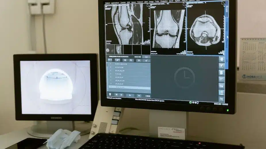
Stretching before a workout to loosen tight muscles. Physical therapy after an injury to facilitate healing. Massage therapy to relieve chronic back pain. These may sound like straightforward treatments, but how can one truly know if they are working under the surface? This uncertainty challenges both patients and therapists. However, emerging ultrasound technology now provides real-time visual feedback to transform treatment precision and efficacy.
What is Real-Time Ultrasound in Therapy?
Real-time ultrasound allows therapists unprecedented visibility into muscles, tendons, ligaments and other tissues beneath the skin. Compact, handheld scanners project sound waves deep into the body and capture the visual reflections. Sophisticated software then translates these images into detailed, moving footage displayed on a screen.
Unlike static MRIs or X-rays, real-time ultrasound shows tissues in motion, contracting and relaxing with natural movements. This real-time feedback helps therapists visually pinpoint and analyze abnormalities. Patients also gain insight into their own bodies, empowering them to actively participate in their recovery.
Advancing Treatment Accuracy with Visualization
Real-time ultrasound eliminates much of the speculation in therapy. Instead of guessing which structures may be impaired based just on symptoms and external exams, visual confirmation guides precise diagnoses. Therapists can then tailor interventions to address the specific dysfunctions revealed.
For example, a patient with knee pain may be experiencing problems with any combination of muscles, tendons, ligaments or cartilage. Traditional manual muscle testing assesses strength and range of motion but provides limited visibility. Ultrasound imaging clearly highlights joint instability or inflammation in the affected structures, enabling targeted treatment.
Such accurate insight also helps patients better understand their own conditions while actively engaging in therapy. Watching targeted tissues respond positively over multiple sessions maintains motivation and compliance. In fact, studies show real-time ultrasound leads to faster recovery, fewer recurring injuries and greater patient satisfaction.
Revolutionary Applications in Rehabilitation Therapy
From stroke rehabilitation to ACL tears in athletes, real-time ultrasound is transforming recovery practices across domains. It serves as an invaluable tool for physical therapists, athletic trainers, chiropractors and other specialists working to restore optimal movement.
For neurological patients, ultrasound feedback helps rebuild neural connections, regain muscle control and retrain balance and coordination. Following a stroke, for instance, visualization of atrophied muscles guides strengthening exercises to mitigate paralysis or limb dysfunction. Patients also rely on the real-time images to recalibrate proper technique.
Sports medicine specialists employ ultrasound technology to accurately evaluate the grade of soft tissue injuries, customize treatment programs and monitor healing without unnecessary downtime. Ultrasound imaging confirms specific tears and also assures both patient and provider when it is truly safe to return to active training or competition without risk of re-injury. This application will likely expand as more therapists recognize the advantages.
A Non-Invasive Approach with Numerous Benefits
Unlike alternatives like MRI, real-time ultrasound provides detailed visualization without high costs or medical risks. There is no exposure to radiation as with X-rays. The technique is also painless and well-tolerated by most patients since there is no need to remain perfectly still for long periods. This makes it ideal for children, pregnant women and elderly individuals concerned about medical procedures.
Portability of modern ultrasound equipment also enables imaging in diverse settings like sports training facilities, physical therapy clinics or even disaster sites when necessary. As technology continues advancing, costs are decreasing to improve accessibility for all patients. Given the multitude of benefits, real-time ultrasound is likely to become standard practice for monitoring and guiding rehabilitation therapy.
Enhancing Treatment of Musculoskeletal Conditions
Chronic neck, shoulder and back pain affect millions globally, diminishing health and quality of life. Real-time ultrasound is at the forefront of accurately diagnosing tricky neuropathic and musculoskeletal discomfort. Detailed images clearly highlight nerve impingement, damaged tendons, muscle tears, herniated discs, arthritis and other common culprits that standard exams struggle to parse out.
Once the origins of patients’ pain are confirmed, therapists can then leverage ultrasound guidance in administering specific exercises, joint manipulation, electrical stimulation or other treatments. They also rely on ultrasound feedback to guarantee proper patient technique and track improvements over time. This level of insight is invaluable in managing complex musculoskeletal conditions. It also builds patient confidence and active participation throughout therapy.
The Future of Therapy Powered by Visualization
As technological capabilities and clinical applications continue advancing, real-time ultrasound promises to expand therapeutic vision and efficacy like never before. Continued software developments even incorporate 3D modeling and artificial intelligence to automatically flag abnormalities and track data over time. Such innovative tools aim to boost treatment insights and consistent quality across the industry.
While mainstream integration across all clinics and specialties will still take some years, the observable benefits make adoption inevitable. In coming decades, visualized therapy powered by real-time ultrasound will likely serve as the ultimate paradigm and gold standard for diverse rehabilitation practices. This marks an exciting shift towards more measurable, efficient and patient-centered therapeutic approaches to improve quality of life.


